
Introduction
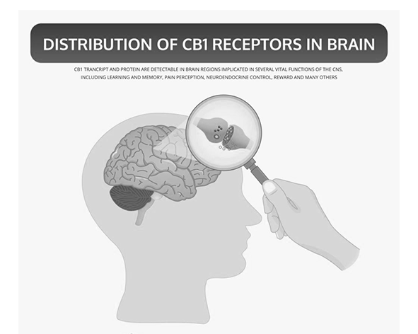
The cannabinoid type 1 receptor (CB1) is a membrane receptor and a core component of the endocannabinoid system. This system combines receptors, lipid messengers and metabolic enzymes to regulate signalling locally. CB1 is particularly abundant in the vertebrate brain, where it can adjust synaptic transmission in circuits involved in memory, movement, nociception and feeding (Herkenham et al., 1990; Kano et al., 2009).
CB1 belongs to the rhodopsin-like, or class A, family of G protein-coupled receptors (GPCRs). Its endogenous ligands include anandamide and 2-arachidonoylglycerol (2-AG). Δ9-tetrahydrocannabinol (THC) also activates it. The effects of receptor activation vary with the ligand, the cell expressing the receptor and the signalling proteins available in that cell.
The same receptor can have very different consequences in different cells. In a nerve terminal, a change in calcium entry can alter the amount of transmitter released by an action potential. In a peripheral cell, coupling to a different set of enzymes and channels can change how that cell responds to other signals. This dependence on cellular context is a recurring theme in CB1 research.
It is useful to distinguish the receptor from the rest of the system. The cell that produces anandamide or 2-AG need not be the cell on which CB1 acts, and the enzymes that remove those lipids determine how long they remain available. Receptor distribution therefore describes where CB1-dependent effects are possible. Understanding a physiological response also requires knowing where the ligand comes from and what happens once the receptor is activated (Lu and Mackie, 2016).
History of discovery
Early explanations of cannabinoid action emphasised the ability of these lipid-soluble molecules to enter cell membranes. A more specific explanation emerged when binding experiments showed a high-affinity site for cannabinoids in rat brain membranes. In 1988, Devane and colleagues characterised that site using a radiolabelled synthetic cannabinoid. Matsuda and colleagues subsequently cloned the receptor and showed that the cloned complementary DNA encoded a functional receptor with seven transmembrane domains (Devane et al., 1988; Matsuda et al., 1990).
Mapping cannabinoid binding sites showed that receptors were distributed unevenly across the brain. Strong signals in the hippocampus, basal ganglia, cerebellum and cortex gave researchers anatomical starting points for studying cannabinoid effects on different circuits (Herkenham et al., 1990).
Cloning CB1 also sharpened a question about normal physiology. What substance in the body binds this receptor? In 1992, anandamide was isolated from brain tissue. In 1995, independent studies identified 2-AG as another endogenous cannabinoid receptor ligand. These discoveries shifted the focus towards a physiological signalling system, including the enzymes that determine when and where the ligands are available.
Radioligand binding and receptor cloning answered different questions. Binding experiments showed that brain membranes contained a site with the expected pharmacological properties. Cloning identified the protein responsible and allowed its activity to be tested in cells expressing the cloned sequence. Anatomical mapping then showed where cannabinoid binding was concentrated. Together, these approaches connected a pharmacological observation to a molecular identity and a set of candidate neural circuits (Devane et al., 1988; Matsuda et al., 1990; Herkenham et al., 1990).
The isolation of endogenous ligands changed the interpretation of those findings. Anandamide, chemically N-arachidonoylethanolamine, and 2-AG are distinct lipid molecules. Their identification opened the way to studying when each is produced, how it acts at the receptor and how its signal is terminated. The modern concept of an endocannabinoid system emerged from this combination of receptor pharmacology and lipid biochemistry (Devane et al., 1992; Mechoulam et al., 1995; Lu and Mackie, 2016).
Molecular structure
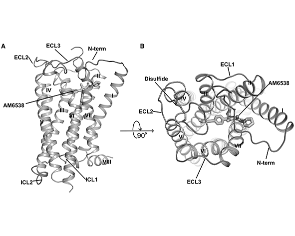
CB1 has seven membrane-spanning helices, designated TM1–TM7, connected by extracellular and intracellular loops. Its amino terminus faces the extracellular space and its carboxyl terminus faces the cytoplasm. This arrangement places ligand recognition and intracellular signalling on opposite sides of a single membrane protein.
Structural studies of human CB1 revealed a largely hydrophobic binding pocket within the transmembrane bundle. Anandamide, 2-AG and THC are lipid-soluble, so partitioning into the membrane can influence how they reach the receptor. Structures captured with different ligands show how the pocket can accommodate chemically distinct molecules and help explain why ligand binding can favour different receptor conformations (Hua et al., 2016; Shao et al., 2016).
On the intracellular side, the loops and the cytoplasmic ends of the helices contribute to G protein coupling. The C-terminal tail also contains regulatory sites that influence what happens after activation, including recruitment of proteins involved in desensitisation and trafficking.
The transmembrane bundle performs two connected jobs. Towards the extracellular side, residues lining the pocket make contacts with the ligand. Towards the cytoplasmic side, the helices and loops form an interface for signalling partners. A ligand can change the relative positions of these elements and thereby affect how readily the receptor engages a G protein. This is the structural basis for distinguishing occupancy of the binding site from activation of a downstream pathway.
High-resolution structures provide snapshots of CB1 in particular experimental states. Crystallography and cryo-electron microscopy can help compare ligand binding and receptor conformations, while functional experiments are needed to establish what those conformations mean in a living cell. The intracellular loops, including the third loop, and the C-terminal region are also important when studying coupling and receptor regulation (Hua et al., 2016; Shao et al., 2016).
Intracellular signalling
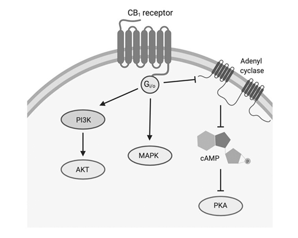
Agonist binding changes the conformation of CB1 and favours coupling to heterotrimeric G proteins, principally of the Gi/o family. One downstream effect is inhibition of adenylyl cyclase, which can lower intracellular cyclic AMP (cAMP) and change the activity of cAMP-dependent pathways.
CB1 also modulates ion channels. Depending on the cell, activation can inhibit N-type and P/Q-type voltage-gated calcium channels and engage G protein-activated inwardly rectifying potassium channels (GIRKs). At a presynaptic terminal, reduced calcium entry can lower the probability that vesicles release neurotransmitter. In other cellular contexts, changes in potassium conductance can affect excitability directly (Kano et al., 2009).
Experimental systems have also linked CB1 to MAPK/ERK, PI3K/Akt and AMPK signalling. The pathways engaged depend on cell type, receptor expression and experimental conditions. The presence of CB1 alone therefore gives limited information about which downstream effect will predominate.
The G protein is itself a source of several signals. Its activated α subunit can influence adenylyl cyclase, while the associated βγ subunits can contribute to ion-channel regulation. A fall in cAMP can alter cAMP-dependent protein activity, while a change in membrane conductance can affect the response on a shorter timescale. These routes can operate together, but the contribution of each depends on which effectors a cell expresses.
At a presynaptic terminal, the sequence matters. Calcium entry normally links electrical activity to vesicle fusion. Inhibiting calcium channels can therefore reduce transmitter release even if action potentials still reach the terminal. The resulting change is a modulation of communication between cells. Its consequences differ at an excitatory terminal and at an inhibitory terminal (Kano et al., 2009).
Endocannabinoid-mediated retrograde signalling
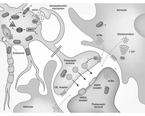
At many synapses, postsynaptic depolarisation increases intracellular calcium, or a postsynaptic receptor activates a pathway that stimulates endocannabinoid synthesis. The cell generates lipid messengers from membrane precursors. In many of the best-characterised examples, 2-AG reaches CB1 on the presynaptic terminal and reduces transmitter release. The direction is retrograde because the signal travels back towards the cell that released the neurotransmitter.
The effect on the circuit depends on the terminal. Suppressing release from a GABAergic terminal briefly reduces inhibition, a phenomenon called depolarisation-induced suppression of inhibition (DSI). Suppressing glutamate release can briefly reduce excitation, termed depolarisation-induced suppression of excitation (DSE). Repeated or sustained patterns of activity can also recruit forms of endocannabinoid-mediated long-term depression (Kano et al., 2009).
The locations of synthetic enzymes and receptors help determine which synapses respond. Ligand breakdown limits the duration of the response. Together, these properties allow a postsynaptic cell to adjust particular inputs without imposing the same change on every nearby terminal.
The postsynaptic trigger can arise in more than one way. Depolarisation can increase intracellular calcium, while activation of certain postsynaptic receptors can recruit lipid-synthetic pathways. In many circuits, diacylglycerol lipase α produces 2-AG from diacylglycerol. The locally produced messenger reaches presynaptic CB1, where Gi/o signalling alters the release machinery. Anandamide can also engage CB1, although the identity and contribution of the lipid messenger depend on the preparation (Kano et al., 2009; Lu and Mackie, 2016).
Duration is another distinction. In DSI and DSE, the suppression after depolarisation is brief. Endocannabinoid-mediated long-term depression involves a sustained decrease in synaptic efficacy following an appropriate pattern of activity. In both cases, the receptor is part of a feedback pathway through which the receiving neuron can influence its own inputs. The spatial arrangement of the synapse and the timing of lipid synthesis and degradation help determine whether the effect remains local and transient.
Regulation and desensitisation
After sustained agonist stimulation, GPCR kinases can phosphorylate residues in the CB1 C-terminal tail. This can favour β-arrestin recruitment and reduce coupling to G proteins. β-Arrestins can also promote receptor internalisation through endocytosis.
Once internalised, CB1 can be recycled to the cell surface or directed towards degradation. Recycling can restore responsiveness, while degradation can reduce the number of receptors available for signalling. The balance between these outcomes depends on the ligand, the duration of stimulation and the cellular machinery that handles the receptor.
Desensitisation and internalisation describe different steps. Desensitisation is a reduction in the ability of a receptor to signal, which can occur while it is still at the cell surface. Internalisation moves the receptor into the cell, where it enters sorting pathways. A receptor that returns to the membrane can again become available for activation. A receptor sent towards lysosomal degradation is removed from the pool that can be recycled.
These processes help explain why a response measured immediately after agonist exposure can differ from one measured after sustained stimulation. The number of receptors at the membrane, their ability to couple to G proteins and the activity of downstream pathways can change on different timescales. The response to a later stimulus therefore depends in part on the cell’s previous exposure.
Biased signalling
Ligands acting on CB1 can produce different relative responses across signalling pathways. A ligand may favour G protein activation over β-arrestin recruitment, or show a different balance between downstream responses. This phenomenon is known as biased signalling.
Assessing bias requires a reference ligand and comparable measurements across pathways. Receptor abundance, signal amplification and the timing of measurements can all change the observed profile. The concept gives researchers a way to test how endogenous, plant-derived and synthetic ligands differ beyond their ability to bind CB1.
A comparison might measure G protein activation and β-arrestin recruitment for two ligands in the same cellular system. If one ligand produces a larger response in both assays, it may simply be more efficacious under those conditions. Evidence for bias concerns the relative balance between the pathways, assessed against a common reference. A result obtained in one assay or cell line should be interpreted in light of that assay’s sensitivity and the available signalling machinery.
For CB1, this distinction is useful because endogenous, plant-derived and synthetic ligands differ in their chemistry and pharmacology. Studying the pathways they engage can clarify how a change at the binding pocket translates into a particular response. The experimental question is which signals a ligand favours under defined conditions and whether the pattern is reproduced in the relevant tissue.
Synaptic organisation and subcellular localisation
Presynaptic CB1 is found on both GABAergic and glutamatergic terminals, with levels and placement varying by brain region and cell type. Its location puts it in a position to modulate transmitter release in response to lipids produced by nearby cells.
Studies have also reported CB1 in dendrites and in intracellular compartments associated with the endoplasmic reticulum and mitochondrial membranes. These observations have prompted work on receptor trafficking and on possible links between intracellular CB1 and cellular metabolism. Detecting the receptor in a compartment and demonstrating its function there are separate experimental questions.
Immunohistochemistry and electron microscopy address different aspects of this organisation. Tissue staining helps identify regions and cell populations with receptor expression. Higher-resolution imaging can help place the receptor relative to particular cellular membranes. At the synapse, that position matters because a receptor near presynaptic release machinery can influence transmission differently from one in a dendrite or intracellular compartment.
The distribution within an individual neuron also helps separate questions about receptor trafficking from questions about signalling. Intracellular CB1 may include receptors being synthesised, transported or internalised. Establishing an additional function for a particular compartment requires evidence that the receptor is present there and that selectively changing its activity changes a relevant cellular response.
Distribution across tissues
CB1 is expressed throughout the central nervous system, but receptor density varies markedly. The hippocampus contains CB1 on inhibitory and excitatory terminals involved in synaptic plasticity. The basal ganglia and cerebellum provide examples in circuits concerned with movement and coordination, while cortical expression is relevant to sensory and cognitive processing (Herkenham et al., 1990; Kano et al., 2009).
CB1 is also found in peripheral neural pathways and in tissues including the liver, adipose tissue, gastrointestinal tract and parts of the reproductive system. Studies in these settings examine lipid metabolism, endocrine signalling and digestive physiology. Anatomical distribution helps explain why CB1 activation can have different consequences in different tissues (Pertwee, 2008; Lu and Mackie, 2016).
In the hippocampus, receptor location on different types of terminal allows endocannabinoids to alter the balance of inhibition and excitation. In the basal ganglia and cerebellum, the same general mechanism operates within circuits organised for movement and coordination. Cortical expression provides another setting, where the effect depends on the local arrangement of excitatory and inhibitory cells. Regional abundance is therefore a starting point for studying function at the level of individual circuits.
Peripheral expression raises a complementary set of questions. In the gastrointestinal tract, investigators can ask how CB1 affects local neural signalling and digestive function. In liver and adipose tissue, studies examine how receptor activity relates to lipid handling and energy balance. In reproductive tissues and endocrine contexts, the relevant cells and signals differ again. Results from the brain cannot be assumed to apply directly to these tissues simply because they express the same receptor (Pertwee, 2008; Lu and Mackie, 2016).
Interactions with phytocannabinoids
THC is a partial agonist at CB1 in many experimental systems, and CB1 mediates much of its psychoactive activity. The response depends on the experimental preparation, including receptor density and how efficiently receptor activation is translated into a downstream signal (Pertwee, 2008).
Cannabidiol (CBD) has relatively low affinity for the orthosteric binding site of CB1. Negative allosteric modulation has been reported in experimental systems, meaning that CBD can alter the receptor’s response to another ligand under the conditions tested. That observation addresses receptor pharmacology. Broader hypotheses about interactions among plant constituents require evidence for the particular combination and outcome being studied.
For lipid-soluble ligands such as THC, the membrane is part of the environment in which binding takes place. The hydrophobic transmembrane pocket can accommodate THC, and receptor activation can then recruit Gi/o-dependent pathways. As with endogenous ligands, the size of the observed response depends on receptor abundance and coupling efficiency in the tissue or assay.
An orthosteric ligand occupies the primary binding site used by an endogenous agonist. An allosteric modulator changes receptor behaviour through a different interaction and can alter the response to a ligand at the primary site. This distinction helps frame experimental reports about CBD. It also separates a specific receptor mechanism from the broader idea of an entourage effect, which concerns possible interactions among plant constituents such as cannabinoids and terpenes.
Biomedical research
CB1 provides a well-characterised model for studying how lipid messengers regulate synapses. Experiments can link receptor location to short-term changes in transmitter release and to longer-term plasticity. Comparing inhibitory and excitatory terminals helps explain why activation of the same receptor can lead to different circuit responses.
Peripheral CB1 is also studied in relation to energy balance and gastrointestinal physiology. Researchers use tissue-specific manipulations and pharmacological tools to distinguish effects mediated by brain circuits from effects originating in peripheral tissues. Structural studies complement this work by linking ligand binding and receptor conformation to measurable cellular responses.
Researchers combine several approaches to connect receptor activity with physiology. Receptor mapping identifies candidate sites of action. Electrophysiology tests how activation affects transmission at identified synapses. Pharmacological and genetic experiments can then test whether the response depends on CB1. Comparing these measurements helps distinguish a direct effect at a terminal from a downstream change elsewhere in the circuit.
Outside the nervous system, the same logic applies to metabolic, endocrine and gastrointestinal research. A tissue may contain CB1, while a whole-organism response can still reflect interactions between that tissue and the brain or other organs. Models that manipulate the receptor in defined cell populations help separate these contributions. Together with structural and pharmacological studies, they provide a way to relate the properties of CB1 to the function of the wider endocannabinoid system.
Figures
References
- Devane WA, Dysarz FA, Johnson MR, Melvin LS, Howlett AC. Identification of a cannabinoid receptor in rat brain. Molecular Pharmacology. 1988.
- Matsuda LA, Lolait SJ, Brownstein MJ, Young AC, Bonner TI. Structure of a cannabinoid receptor and functional expression of the cloned cDNA. Nature. 1990.
- Herkenham M, Lynn AB, Little MD, et al. Cannabinoid receptor localization in brain. Proceedings of the National Academy of Sciences. 1990.
- Devane WA, Hanus L, Breuer A, et al. Isolation and structure of a brain constituent that binds to the cannabinoid receptor. Science. 1992.
- Mechoulam R, Ben-Shabat S, Hanus L, et al. Identification of an endogenous 2-monoglyceride present in canine gut that binds to cannabinoid receptors. Biochemical Pharmacology. 1995.
- Di Marzo V, Fontana A. Anandamide and other endocannabinoids: metabolism and biological functions. Prostaglandins, Leukotrienes and Essential Fatty Acids. 1995.
- Di Marzo V, Bifulco M, De Petrocellis L. The endocannabinoid system and its therapeutic exploitation. Nature Reviews Drug Discovery. 2004.
- Pertwee RG. The diverse CB1 and CB2 receptor pharmacology of cannabinoids. British Journal of Pharmacology. 2008.
- Kano M, Ohno-Shosaku T, Hashimotodani Y, Uchigashima M, Watanabe M. Endocannabinoid-mediated control of synaptic transmission. Physiological Reviews. 2009.
- Lu HC, Mackie K. An introduction to the endogenous cannabinoid system. Biological Psychiatry. 2016.
- Hua T, Vemuri K, Pu M, et al. Crystal structure of the human CB1 cannabinoid receptor. Cell. 2016.
- Shao Z, Yin J, Chapman K, et al. High-resolution crystal structure of the human CB1 cannabinoid receptor. Nature. 2016.