
Introduction
The endocannabinoid system (ECS) is a network of lipid signals found across vertebrate tissues. Its core components are G protein-coupled cannabinoid receptors, endogenous ligands derived from polyunsaturated fatty acids, and enzymes that synthesise and break down those ligands. Together, they allow cells to adjust their activity in response to local conditions and contribute to physiological homeostasis (Di Marzo, 2008; Lu and Mackie, 2016).
The system was discovered through research on Δ9-tetrahydrocannabinol (THC), the principal psychoactive cannabinoid in Cannabis sativa. The identification of specific binding sites in rat brain raised the question of what normally activated those receptors. The discovery of CB2 in peripheral tissues and the isolation of anandamide and 2-arachidonoylglycerol (2-AG) established that cannabinoid receptors form part of an endogenous signalling system.
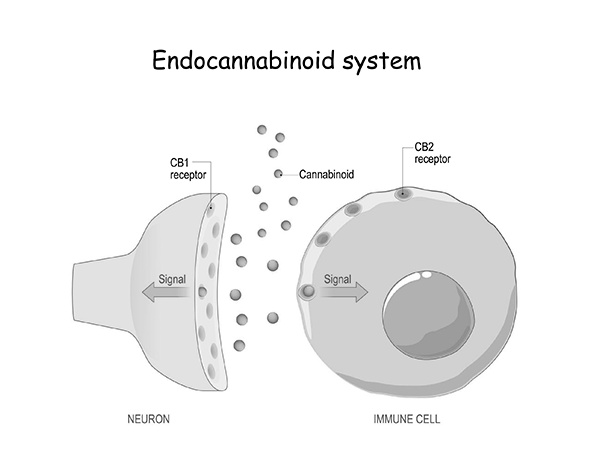
Many conventional neurotransmitters are stored in presynaptic vesicles. At the synapses where endocannabinoid signalling is best characterised, cells instead make these lipid messengers from membrane precursors in response to a stimulus. The messengers act locally and can travel back across a synapse to regulate transmitter release. This mechanism is particularly well studied in the nervous system, while other forms of endocannabinoid signalling operate in immune, metabolic and endocrine tissues.
History of discovery
Identification of the cannabinoid receptors
For much of the twentieth century, the high lipid solubility of cannabinoids made it plausible that their effects arose from non-specific interactions with cell membranes. Pharmacological evidence challenged that account. The effects of cannabinoids showed stereoselectivity, and binding could be saturated, both consistent with interaction at a specific receptor.
In 1988, Devane and colleagues used radioligand binding to identify a high-affinity cannabinoid binding site in rat brain membranes. Matsuda and colleagues subsequently cloned the receptor now known as CB1 and showed that its complementary DNA encoded a functional receptor with seven transmembrane domains. Autoradiographic studies mapped cannabinoid binding across the rat brain and found marked differences between regions, including the hippocampus, basal ganglia, cerebellum and cerebral cortex (Devane et al., 1988; Matsuda et al., 1990; Herkenham et al., 1990).
In 1993, Munro and colleagues characterised CB2. Its association with immune cells and peripheral tissues extended the study of cannabinoid signalling beyond the brain. The identification of two receptors made the search for naturally occurring ligands a concrete experimental question.
Discovery of the endocannabinoids
In 1992, Devane and colleagues isolated N-arachidonoylethanolamine from pig brain extracts. The compound became known as anandamide (AEA), from the Sanskrit ānanda, meaning bliss or well-being. It binds CB1 and reproduced several effects observed with plant-derived cannabinoids in experimental systems (Devane et al., 1992).
Two independent groups reported a second endogenous cannabinoid receptor ligand, 2-AG, in 1995. In many tissues, 2-AG is present at higher concentrations than anandamide. The two ligands also differ pharmacologically. Anandamide can act as a partial agonist at CB1, whereas 2-AG is generally characterised as a full agonist at both CB1 and CB2 in experimental assays (Mechoulam et al., 1995; Sugiura et al., 1995; Lu and Mackie, 2016).
From receptors and ligands to a signalling system
Characterising fatty acid amide hydrolase (FAAH) and monoacylglycerol lipase (MAGL) helped explain how cells terminate the signals carried by anandamide and 2-AG. The resulting model describes a dynamic system in which the availability of a ligand depends on both how quickly it is produced and how quickly it is cleared (Cravatt et al., 1996; Dinh et al., 2002).
Components of the endocannabinoid system
The core system comprises cannabinoid receptors, their endogenous lipid ligands and enzymes that control ligand synthesis and degradation. Transient production from membrane lipids and subsequent enzymatic breakdown give cells several ways to regulate a signal locally.
Cannabinoid receptors
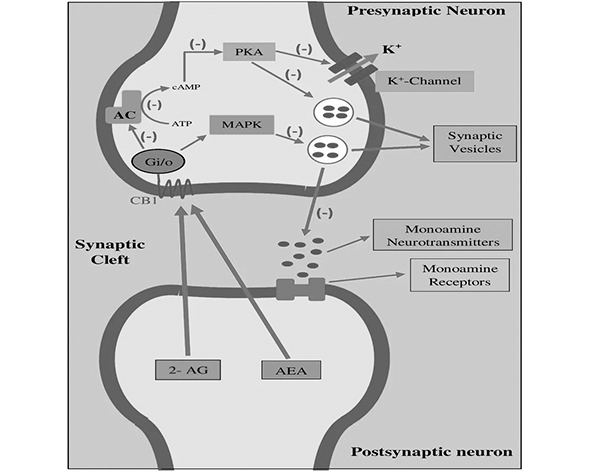
CB1 and CB2 are seven-transmembrane G protein-coupled receptors (GPCRs). They commonly couple to Gi/o proteins. Their activation can inhibit adenylyl cyclase and alter downstream signalling, with the consequences depending on the cell and its complement of signalling proteins (Howlett et al., 2002).
CB1
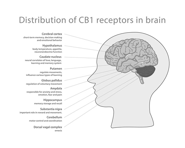
CB1 is among the most abundant GPCRs in the central nervous system. It is expressed in the hippocampus, cerebellum, basal ganglia, cerebral cortex and amygdala, among other regions. CB1 activation can lower intracellular cyclic AMP, modulate voltage-dependent ion channels and engage pathways such as MAPK/ERK. At presynaptic terminals it often reduces transmitter release. Depending on the circuit, the transmitters affected include glutamate, GABA, dopamine and acetylcholine (Herkenham et al., 1990; Mackie, 2008).
CB2
CB2 was first characterised as a peripheral cannabinoid receptor. It is associated with haematopoietic tissues and immune populations including macrophages and B and T lymphocytes. Expression has also been reported in some nervous-system cells, including microglia. Abundance can vary with cell type and activation state, an important distinction when interpreting findings from different tissues or experimental conditions (Munro et al., 1993; Lu and Mackie, 2016).
Endocannabinoids
Anandamide (AEA)
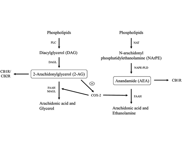
Anandamide is also known as N-arachidonoylethanolamine. One route to its synthesis uses N-acylphosphatidylethanolamines (NAPEs) as precursors and involves NAPE-specific phospholipase D (NAPE-PLD). Other synthetic routes exist. Anandamide is generally less abundant than 2-AG in tissue measurements, but its activity depends on local availability and the targets present. In addition to cannabinoid receptors, it can engage the TRPV1 ion channel under some conditions.
2-Arachidonoylglycerol (2-AG)
2-AG is abundant in the brain and many peripheral tissues. Diacylglycerol lipases DAGLα and DAGLβ generate it from diacylglycerol. In many synapses it is the main messenger that reaches presynaptic CB1 after postsynaptic activity. It can activate CB2 as well as CB1 (Sugiura et al., 1995; Kano et al., 2009; Lu and Mackie, 2016).
Enzymes that terminate or modify the signal
FAAH. Fatty acid amide hydrolase hydrolyses anandamide to arachidonic acid and ethanolamine. Its association with intracellular membranes, including the endoplasmic reticulum, helps determine where anandamide is metabolised (Cravatt et al., 1996).
MAGL. Monoacylglycerol lipase hydrolyses 2-AG to arachidonic acid and glycerol. Biochemical profiling attributed approximately 85% of 2-AG hydrolytic activity in the mouse brain preparations studied to MAGL. The estimate describes those experimental preparations and need not apply uniformly across tissues (Dinh et al., 2002; Blankman et al., 2007).
Other enzymes. ABHD6 and ABHD12 also contribute to 2-AG hydrolysis. COX-2 can metabolise endocannabinoids through oxidation. These routes help explain why the enzymes controlling local lipid concentrations can differ between cells and physiological conditions.
Mechanisms of endocannabinoid signalling
Retrograde signalling at synapses
At many synapses, the postsynaptic cell can send a signal back to the terminal that released the neurotransmitter. The sequence can be described in four steps (Wilson and Nicoll, 2001; Kano et al., 2009; Castillo et al., 2012).
- Postsynaptic depolarisation raises intracellular calcium, or a postsynaptic GPCR activates a pathway that promotes endocannabinoid synthesis.
- The cell makes lipid messengers from membrane precursors. DAGLα generates 2-AG in many of the best-characterised circuits.
- The messenger reaches presynaptic CB1 and activates Gi/o signalling.
- Presynaptic signalling lowers the probability of neurotransmitter release, for example by reducing calcium entry through voltage-dependent channels.
Depolarisation-induced suppression
Depolarisation-induced suppression of inhibition (DSI) is a transient reduction in inhibitory input after postsynaptic depolarisation, typically involving reduced GABA release. Depolarisation-induced suppression of excitation (DSE) is the corresponding reduction in excitatory input, typically involving glutamate. The same feedback mechanism can therefore have different effects on a circuit depending on which input is suppressed.
Synaptic plasticity
Endocannabinoid signalling can also produce longer-lasting changes in synaptic strength. In endocannabinoid-mediated long-term depression (eCB-LTD), a pattern of activity recruits CB1 and leads to a sustained reduction in synaptic efficacy. Forms of this plasticity have been studied in the hippocampus, prefrontal cortex, striatum and cerebellum. They provide experimental models for investigating learning, memory and neuronal adaptation (Kano et al., 2009; Castillo et al., 2012).
Spatial and temporal control
The location of synthetic enzymes, the position of receptors and the rate of lipid breakdown determine how far a signal spreads and how long it lasts. FAAH primarily clears anandamide, while MAGL accounts for much of the 2-AG hydrolysis in the brain. ABHD6 and ABHD12 make additional contributions. This arrangement allows neighbouring synapses to respond differently to the same activity (Blankman et al., 2007).
Functional significance
By regulating both inhibitory and excitatory input, endocannabinoids help shape neuronal excitability. Experimental studies have examined this modulation in circuits involved in reward, emotional processing, learning, memory and nociception. The effects depend on the circuit and on the synapses expressing CB1.
Distribution across tissues
Cannabinoid receptors and endocannabinoid metabolic enzymes have been identified in many organ systems. The high density of CB1 in the brain first drew attention to neuronal signalling, but peripheral tissues can also make, respond to and metabolise endocannabinoids (Di Marzo, 2008; Lu and Mackie, 2016).
Central nervous system
Receptor mapping shows uneven CB1 distribution across the brain. Prominent regions include the hippocampus, associated with learning and memory, the cerebellum and basal ganglia, involved in movement and coordination, the cerebral cortex, and the amygdala, which participates in emotional processing. CB2 has also been reported in some central nervous system cells, particularly glial populations such as microglia, where it has been studied in neuroimmune contexts (Herkenham et al., 1990; Mackie, 2008).
Peripheral nervous system
Endocannabinoid components are found in sensory neurons and autonomic pathways. Here they can influence the transmission of nociceptive signals, the processing of other sensory information from peripheral tissues and the regulation of autonomic reflexes.
Immune system
CB2 expression has been described in B and T lymphocytes, macrophages, dendritic cells and natural killer cells. Experimental studies have examined how CB2 activity affects cell migration, cytokine release and inflammatory signalling. The response varies with the immune population and its activation state (Klein, 2005).
Metabolic tissues
The liver, adipose tissue, skeletal muscle and pancreas contain components of the ECS. Research has examined how local signalling relates to energy balance, lipid metabolism and insulin sensitivity. Studies in mice have also linked CB1 to central control of feeding and to lipid synthesis in peripheral tissues (Cota et al., 2003).
Digestive system
Components of the ECS are found in enteric neurons, intestinal epithelial cells and sensory endings in the gastrointestinal tract. Their distribution has prompted research into intestinal motility, gastrointestinal secretion and visceral sensation. The physiological effect depends on where the signal originates and which cells receive it.
Endocrine and reproductive systems
Endocannabinoid components have been described in the hypothalamus, pituitary and gonads. Studies in these tissues examine interactions with hormonal pathways involved in endocrine secretion, reproduction and early development. Tissue expression helps identify where such interactions could occur, while functional experiments are needed to establish their role.
Why tissue distribution matters
Different tissues can use the same lipid messenger in different ways. Receptor expression, local synthesis and enzyme activity determine which cells respond and when. The widespread distribution of these components gives endocannabinoid signalling a role in coordinating neural, immune and metabolic responses.
Physiological functions and homeostasis
Homeostasis involves keeping internal conditions within a workable range as the environment and the organism change. Endocannabinoid signalling provides feedback at the level of individual cells and circuits. Its physiological consequences depend on how that local feedback interacts with other regulatory systems (Di Marzo, 2008; Lu and Mackie, 2016).
Regulation of neurotransmission
Presynaptic CB1 can reduce the release of glutamate or GABA, modifying excitatory or inhibitory input. Endocannabinoid signalling can also affect pathways involving dopamine, which participates in reward and motivation, and acetylcholine, which has cognitive and neuromuscular roles. The net effect depends on the organisation of the circuit and the inputs affected (Kano et al., 2009).
Regulation of energy metabolism
CB1 is expressed in brain regions involved in feeding, including the hypothalamus. Peripheral tissues such as the liver, adipose tissue and skeletal muscle also contain endocannabinoid components. Animal studies distinguish central effects on food intake from peripheral effects on lipid synthesis and investigate how these pathways relate to energy storage and glucose utilisation (Cota et al., 2003).
Regulation of immune responses
In immune cells, CB2 signalling can influence cytokines and other inflammatory mediators. Its effects depend on the cell type and the stimulus. Experimental work in lymphocytes, macrophages and dendritic cells helps distinguish effects on individual cells from changes in the broader inflammatory response (Klein, 2005).
Stress and physiological adaptation
Experimental research implicates endocannabinoid signalling in stress-sensitive circuits involving the amygdala, hippocampus and prefrontal cortex, as well as in regulation of the hypothalamic–pituitary–adrenal axis. The contribution of a ligand can vary with brain region and with the type, timing and duration of the stressor (Hill et al., 2010).
Integration across systems
Neural, immune and metabolic responses often occur together. The distribution of receptors and enzymes allows endocannabinoid signals to help adjust these responses in parallel, while the mechanisms remain specific to individual cells and tissues.
Biomedical research
Neuroscience
The abundance of CB1 and the accessibility of retrograde signalling to experimental study have made synapses a major focus of ECS research. Work in the hippocampus, cortex and amygdala examines how short-term changes in transmitter release and longer-term plasticity relate to learning, memory, emotional processing and nociception (Kano et al., 2009; Castillo et al., 2012).
Immunology
Studies of CB2 investigate how receptor activity changes the behaviour of lymphocytes, macrophages and dendritic cells. These experiments help identify which lipid signals affect cell migration or inflammatory mediators, and how the response changes when cells become activated (Klein, 2005).
Metabolism and energy balance
Researchers distinguish actions in brain circuits that regulate feeding from actions in peripheral tissues that handle lipids and glucose. Comparing receptor activity across tissues and animal models helps establish which mechanisms account for changes in overall energy balance (Cota et al., 2003).
Molecular complexity and the endocannabinoidome
Several cannabinoid-related lipids can interact with targets beyond CB1 and CB2, including TRP ion channels, nuclear receptors such as PPARs and additional GPCRs. They can also share enzymes and precursors with other lipid pathways. The term endocannabinoidome describes this wider network of related lipids, enzymes and molecular targets (Di Marzo and Piscitelli, 2015).
Future directions
Open questions include which lipid mediators are produced under particular physiological conditions, how their signalling interacts with other cellular pathways, and which enzymes control synthesis and breakdown in specific tissues. Relating molecular measurements to functional experiments remains essential for explaining how local signals contribute to whole-organism physiology.
Figures
References
- Blankman JL, Simon GM, Cravatt BF. (2007). A comprehensive profile of brain enzymes that hydrolyze the endocannabinoids 2-arachidonoylglycerol and anandamide. Chemistry & Biology.
- Castillo PE, Younts TJ, Chávez AE, Hashimotodani Y. (2012). Endocannabinoid signaling and synaptic function. Neuron.
- Cota D, Marsicano G, Tschöp M, et al. (2003). The endogenous cannabinoid system affects energy balance via central orexigenic drive and peripheral lipogenesis. Journal of Clinical Investigation.
- Cravatt BF, Giang DK, Mayfield SP, et al. (1996). Molecular characterization of an enzyme that degrades neuromodulatory fatty-acid amides. Nature.
- Devane WA, Dysarz FA, Johnson MR, Melvin LS, Howlett AC. (1988). Determination and characterization of a cannabinoid receptor in rat brain. Molecular Pharmacology.
- Devane WA, Hanus L, Breuer A, et al. (1992). Isolation and structure of a brain constituent that binds to the cannabinoid receptor. Science.
- Di Marzo V. (2008). Targeting the endocannabinoid system: to enhance or reduce? Nature Reviews Drug Discovery.
- Di Marzo V, Piscitelli F. (2015). The endocannabinoid system and its modulation by phytocannabinoids. Neurotherapeutics.
- Dinh TP, Carpenter D, Leslie FM, et al. (2002). Brain monoglyceride lipase participating in endocannabinoid inactivation. PNAS.
- Herkenham M, Lynn AB, Johnson MR, et al. (1990). Characterization and localization of cannabinoid receptors in rat brain. Proceedings of the National Academy of Sciences.
- Hill MN, Patel S, Campolongo P, Tasker JG, Wotjak CT, Bains JS. (2010). Functional interactions between stress and the endocannabinoid system: from synaptic signaling to behavioral output. Journal of Neuroscience.
- Howlett AC, Barth F, Bonner TI, et al. (2002). International Union of Pharmacology. Classification of cannabinoid receptors. Pharmacological Reviews.
- Kano M, Ohno-Shosaku T, Hashimotodani Y, Uchigashima M, Watanabe M. (2009). Endocannabinoid-mediated control of synaptic transmission. Physiological Reviews.
- Klein TW. (2005). Cannabinoid-based drugs as anti-inflammatory therapeutics. Nature Reviews Immunology.
- Lu HC, Mackie K. (2016). An introduction to the endogenous cannabinoid system. Biological Psychiatry.
- Mackie K. (2008). Cannabinoid receptors: where they are and what they do. Handbook of Experimental Pharmacology.
- Matsuda LA, Lolait SJ, Brownstein MJ, Young AC, Bonner TI. (1990). Structure of a cannabinoid receptor and functional expression of the cloned cDNA. Nature.
- Mechoulam R, Ben-Shabat S, Hanus L, et al. (1995). Identification of an endogenous 2-monoglyceride cannabinoid receptor agonist. Biochemical Pharmacology.
- Munro S, Thomas KL, Abu-Shaar M. (1993). Molecular characterization of a peripheral receptor for cannabinoids. Nature.
- Sugiura T, Kondo S, Sukagawa A, et al. (1995). 2-Arachidonoylglycerol: a possible endogenous cannabinoid receptor ligand in brain. Biochemical and Biophysical Research Communications.
- Wilson RI, Nicoll RA. (2001). Endogenous cannabinoids mediate retrograde signalling at hippocampal synapses. Nature.